Tricuspid Clip: Difference between revisions
From Bay Area Structural Heart Wiki
No edit summary |
No edit summary |
||
| Line 1: | Line 1: | ||
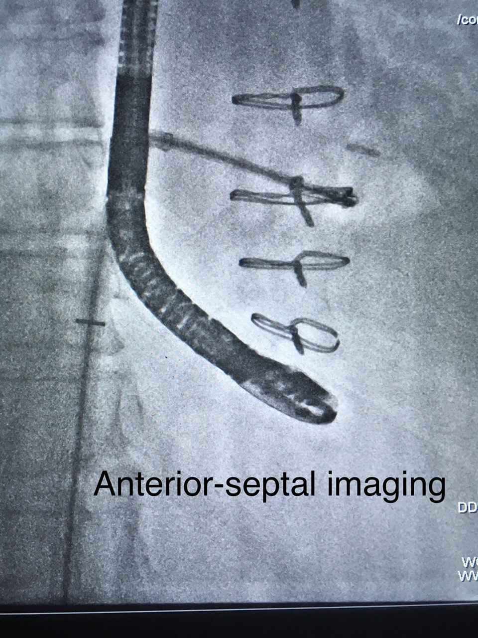
[[File:AntSeptTricuspidClip.jpeg |thumb|150px|right|Anterior Septal imaging for Tricuspid clip|link={{filepath: AntSeptTricuspidClip.jpeg}}]] | [[File:AntSeptTricuspidClip.jpeg|thumb|150px|right|Anterior Septal imaging for Tricuspid clip|link={{filepath:AntSeptTricuspidClip.jpeg}}]] | ||
Revision as of 17:14, 14 August 2020
DRAFT
| Tricuspid Clip | |||||
|---|---|---|---|---|---|
| Anesthesia | Imaging | Access | Pre-Procedure | ||
| General |
|
4F Venous sheath LFV for ACT's and potential ICE access |
| ||
Big Equipment:
Open
- Cath pack
- Lift / Footstool / Support base
- Clear Plexiglass base support
- Sterile System Stabilizer
Standby
- ICE 8fr. and SwiftLink cover
- 9fr x 23cm Pinnacle for ICE
Wires:
Open
- Amplatz SS 7cm
Standby
- 150cm J wire
Misc supplies:
Open
- (4) 108cm High pressure IV Lines
- (8) Stopcocks
- 8F Sheath for dilator - (also used as a backup sheath)
- (1)Perclose
- 4-0 Vicryl/monocryl
- Pickups (Either from a suture removal kit or individual from SPD)
- (2) 60cc syringe
- 12” extension (Keep stickers to label flush tubing)
- Sterile towel pack for back table
- Fluoro cover to anesthesia IV pole or moving xray shield
- 20F Dilator
- Micropuncture
- U/S probe cover
Standby
- Additional Perclose
- Pericardiocentesis supplies
Non-Sterile Supplies
- (4) Liter bags of heparinized saline
- (4) Liter sized pressure bags
Procedure
- Anesthesia will obtain radial art line prior to draping.
- Pre procedure TEE imaging performed
- Prior to prepping patient, place MitraClip Plate (plexiglass board – hinge side up) under patient’s leg off center to the right. Place MitralClip Lift (step stool) over patients right leg, angled down towards the head. Measure 80cm from subxyphoid, or 85 from mid-sternum to leading edge of stool. Make sure there is direct contact between stool and plate.
- Prep patient and drape per usual. Drape anesthesia pole and/or xray shield.
- Obtain RFV access, dilate with 8F, preclose. .035 wire inserted into perclose.
- Amplatz SS advanced through perclose to RV
- 20F dilator inserted and removed
- MitraClip Guide inserted.
- Setup stabilizer and silicone sheet. Hand MD 60ml syringe with short extension tubing for aspiration.
- Amplatz wire and dilator removed. Clip inserted.
- Drips should be maintained at 1 drip every 3 seconds. If bubbles are seen on echo, turn drips down.
- When clip is being deployed turn room lights up and drips up. Place bowl below to catch drips while deploying.
- Hand MD 60ml syringe with extension tubing for aspiration while delivery system is removed.
- Vicryl used for groin closure.
BACK TABLE SETUP
- 108" Tubing spikes handed off to circulator, keep rollers on sterile field.
- Follow Rep instructions for device prep.
APPROVED: MD initials MM/YY